Comparative Analysis and Evolution Assessment of Dermascopic Images
Featured in:
MD Thesis
Authors:
Tadeu Ferreira
Abstract
According to statistics, Skin Cancer represents one of the most prevalent cancers worldwide with a rapid increase in the last decades. Within skin cancer, Melanoma represents the most dangerous causing more deaths than any other cancer. Fortunately, early diagnosis of Melanoma has proven to be curable making the regular monitoring of the skin essential. When monitoring the skin, dermatologist looks for patterns changes in previously pigmented skin lesions. However, quantification of patterns over time is a difficult task even for the most experienced doctor.Over the years, many automated diagnostic systems have been developed for classification of malignant lesions. However, there has been very little work directed towards the assessment of the evolution of pigmented skin lesions. Development of an automated system which detects a significant evolution of the skin lesion can be used as an additional tool by providing a warning to dermatologists and patients. This study investigates the assessment of the evolution of a skin lesion through comparative analysis of dermoscopic images acquired over time. Special attention has also been paid off how the acquisition of dermoscopic images may influence the assessment of evolution. The assessment of evolution was performed in a two-step algorithm. The first step consisted of automated segmentation of the lesion area in the two images being compared. Afterward, alignment of the binary mask was performed in order to extract border and shape features which could relate to an evolution of the lesion. The assessment was computed with the clustering methods Fuzzy C-Means and Gaussian Mixture Models and, Decision Tree Classification. The performance was evaluated using labels extracted from multiple annotators.The results suggested that assessing the evolution with only border and shape features is insufficient. This study also indicates that an incorrect acquisition of the dermoscopic images would have negative impacts on the correct extraction of features. An analysis has also been conducted that shows that the extraction of labels from annotators with no medical experience affects the algorithm performance which limited the assessment of the evolution of the skin lesion. The results of the assessment of evolution are presented such that they can be used as an incentive to attract more research in this area of study.
Citation
Tadeu Ferreira (2018), Comparative Analysis and Evolution Assessment of Dermascopic Images. MD Thesis. University of Coimbra, 2018.
FLOWING: Implicit Neural Flows for Structure-Preserving Morphing
Authors: Arthur Bizzi; Matias Grynberg; Vitor Matias; Daniel Perazzo; João Paulo Lima; Luiz Velho; Nuno Gonçalves; João Pereira; Guilherme Schardong; Tiago Novello
Featured in: 39th Conference on Neural Information Processing Systems (NeurIPS 2025)
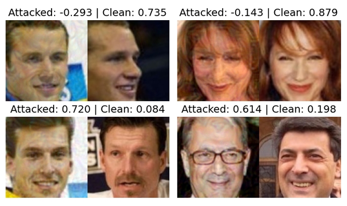
Adversarial Attack Challenge for Secure Face Recognition 2025
Authors: João Tremoço, Iurii Medvedev, Nuno Freitas, Andreia Costa, Diogo Nunes, Niklas Bunzel, Lukas Graner, Nicholas Göller, Lorenzo Pellegrini, Nicolò Di Domenico, Guido Borghi, Monson Verghese, Shruti Bhilare, Avik Hati, Miguel Lourenço, Nuno Gonçalves
Featured in: IEEE International Joint Conference on Biometrics (IJCB 2025)
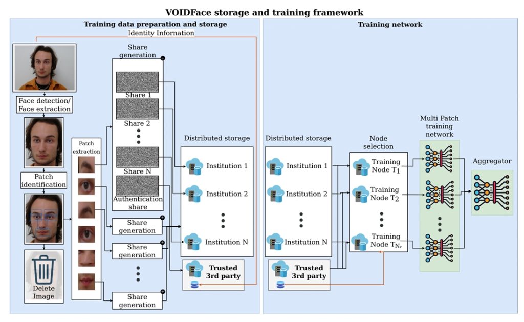
VOIDFace: A Privacy-Preserving Multi-Network Face Recognition With Enhanced Security