Graph-Based Radiomics Feature Extraction from 2D Retina Images
Featured in:
IEEE Access
Authors:
Ofélio Jorreia; Nuno Gonçalves; Rui Cortesão
Abstract
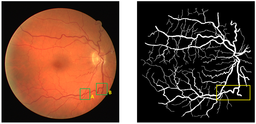
Medical image analysis offers valuable visual support for clinical decision-making, yet the incorporation of quantitative data is essential for deeper diagnostic insight. The radiomics approach addresses this need by combining quantitative image analysis with Machine Learning (ML) techniques, further enhancing Explainable Artificial Intelligence (XAI) for clinical applications. While working with two-dimensional (2D) images derived from volumetric data offers computational advantages, accurately estimating structural properties within these images remains challenging. Within the radiomics framework, this study introduces a methodology to distinguish bifurcations from other structural variations in 2D local fragments of retinal vasculature. Using a publicly available dataset of 29 retinal images, we extracted 1003 feature fragments for experiments. The regions of interest (ROIs) are identified using morphological image processing techniques. Specifically, candidate points are detected by applying structuring elements (SEs) to the skeletonized and binarized vasculature. From each candidate point, a local fragment of 35×35 pixels is extracted and used as input to the classification model. A Convolutional Neural Network (CNN) model, tailored for small image datasets and binary classification tasks is created. The trained model achieved an accuracy of 94.95% in correctly identifying bifurcation points. Based on predicted bifurcation points and blood vessel segments, we use the Graph-Based Radiomics Feature Extraction Algorithm (Graph-BRFExtract) to extract the adjacency matrix. This matrix serves as mathematical representation of the retinal vascular network, constituting a novel form of graph-based radiomic features.
Citation
Ofélio Jorreia, Nuno Gonçalves and Rui Cortesão. Graph-Based Radiomics Feature Extraction from 2D Retina Images. IEEE Access. DOI: 10.1109/ACCESS.2025.3588817.
FLOWING: Implicit Neural Flows for Structure-Preserving Morphing
Authors: Arthur Bizzi; Matias Grynberg; Vitor Matias; Daniel Perazzo; João Paulo Lima; Luiz Velho; Nuno Gonçalves; João Pereira; Guilherme Schardong; Tiago Novello
Featured in: 39th Conference on Neural Information Processing Systems (NeurIPS 2025)
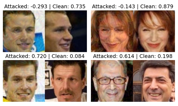
Adversarial Attack Challenge for Secure Face Recognition 2025
Authors: João Tremoço, Iurii Medvedev, Nuno Freitas, Andreia Costa, Diogo Nunes, Niklas Bunzel, Lukas Graner, Nicholas Göller, Lorenzo Pellegrini, Nicolò Di Domenico, Guido Borghi, Monson Verghese, Shruti Bhilare, Avik Hati, Miguel Lourenço, Nuno Gonçalves
Featured in: IEEE International Joint Conference on Biometrics (IJCB 2025)
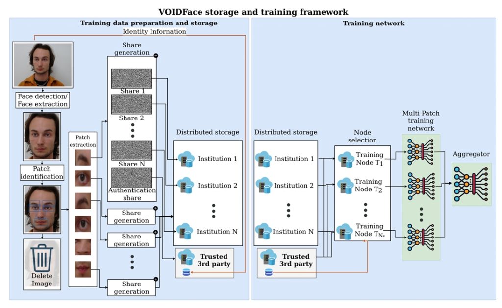
VOIDFace: A Privacy-Preserving Multi-Network Face Recognition With Enhanced Security